Practice Essentials
Interphalangeal (IP) joint dislocations of the fingers and toes are common. [1, 2, 3] Typically associated with forced hyperextension or hyperflexion of the digit, they require immediate reduction. The IP joint is a hinge joint that allows only flexion and extension and consists of several ligamentous complexes. The volar plate provides stability against hyperextension injury and dorsal dislocation of the phalanx. It often ruptures during a dorsal dislocation and may be associated with an avulsion fracture at the base of the phalanx. The strong collateral ligament complex resists hyperextension and lateral dislocation injury. The extensor hood complex stabilizes against hyperflexion injury and volar displacement of the phalanx.
Dislocations of the distal IP (DIP) joint of the fingers are often associated with fracture, tendon rupture, and/or proximal interphalangeal (PIP) joint involvement. Upon axial loading and hyperextension of the fingertip, the volar plate of the DIP joint tears and the joint may be displaced. [4, 5, 6, 7] Forced hyperflexion results in a volar IP joint dislocation (eg, where the distal phalanx is dislocated volar to the middle phalanx). [8]
According to studies, 59.4% of all dislocations involve the joints of the thumb or little finger, with the highest dislocation rates being in the proximal interphalangeal joint of the little finger, the metacarpophalangeal joint of the thumb, and the proximal interphalangeal joint of the ring finger. Dislocations are relatively uncommon in children. Lateral bending forces are more often transmitted through the physis rather than the collateral ligaments in a child‘s hand because the growth plate is the path of least resistance. Although rare, the thumb metacarpophalangeal (MCP) joint is the most commonly dislocated joint in the skeletally immature hand. The PIP joint is the most commonly injured articular surface, involving volar plate or collateral ligament avulsion fractures. [9, 10]
Causes of interphalangeal dislocations include axial compression or lateral forces directed to the digit; forced hyperextension or hyperflexion of the digit from traumatic athletic injury, entrapment of finger between objects, or a fall; and predisposition to ligamentous injury in those with lax ligaments (eg, Down syndrome).
Diagnosis
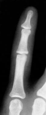
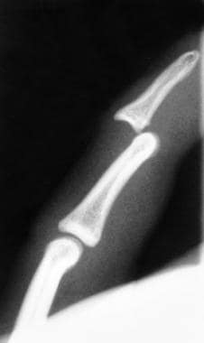
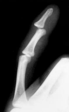
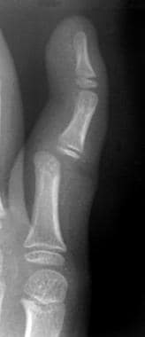
Take anteroposterior, true lateral, and oblique radiographs of the affected digit. Obtain 3 views prior to and after reduction.
(See the images below.)
Treatment
Splint, ice, and elevate the affected digit. [11, 12] Neurovascular status should be evaluated before and after transport to the ED. Administer digital block anesthesia 10-15 minutes before any reduction maneuver. Be sure to remove all rings.
With the patient's hand or foot securely braced, grasp the dislocated phalanx with dry gauze loosely wrapped around the phalanx, and hyperextend the joint slightly with gentle longitudinal traction for a dorsal dislocation or hyperflex for a volar dislocation. Gradually push the dislocated phalanx into its normal anatomic position. [11, 12] Do not apply vigorous traction in a child, because that may interpose soft tissue or an osteochondral fragment into the distracted joint space and prevent reduction. After reduction, examine the affected joint for flexor-extensor tendon function, active range of motion, localized tenderness, and instability in the medial-lateral and dorsal-volar directions. Immobilize the joint with a foam-padded splint immediately after reduction to prevent redislocation or instability.
Joint instability or neurovascular compromise after reduction requires immediate orthopedic or hand consultation. Because joint instability or dysfunction and subtle ligamentous, cartilaginous, or bony injury often are obscured by extensive edema and pain immediately after the injury, all finger joint dislocations should be referred for orthopedic or hand specialist evaluation within 2-3 weeks following reduction.
If joint reduction by means of closed manipulation is not feasible due to interposition of periarticular soft tissues, an irreducible dislocation is diagnosed. Irreducible dislocations of the proximal interphalangeal joint are rare injuries. However, every physician should recognize these injuries and immediately refer them to a hand surgeon. [13]
Patients whose digits have neurovascular compromise, an open joint dislocation, ligamentous or volar plate rupture, joint instability, or an associated fracture should have immediate orthopedic consultation. All finger dislocations should be reevaluated subsequently by an orthopedic or hand specialist to manage potential subtle ligamentous, cartilaginous, or bony injury. [11, 12] A lateral or volar PIP joint dislocation, although rare, requires an orthopedist for possible open reduction with internal fixation. A dislocation of the metacarpophalangeal (MCP) joint, although rare in adults, may be more common in children. [14, 15, 16] MCP dislocation usually requires open reduction by a pediatric orthopedist.
Also see Joint Reduction, Finger Dislocation and Joint Reduction, Thumb Dislocation.
-
Anteroposterior view of distal interphalangeal (DIP) joint dislocation
-
Lateral view of distal interphalangeal (DIP) joint dislocation
-
Oblique view of distal interphalangeal (DIP) joint dislocation
-
Oblique view of proximal interphalangeal (PIP) joint dislocation


